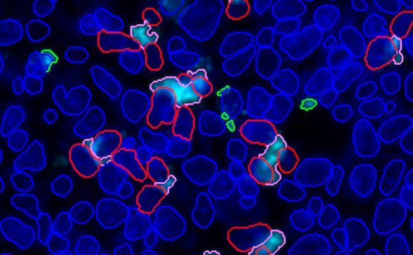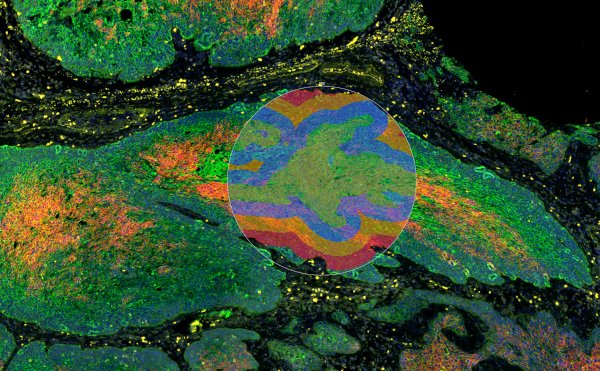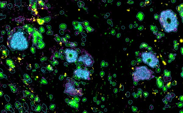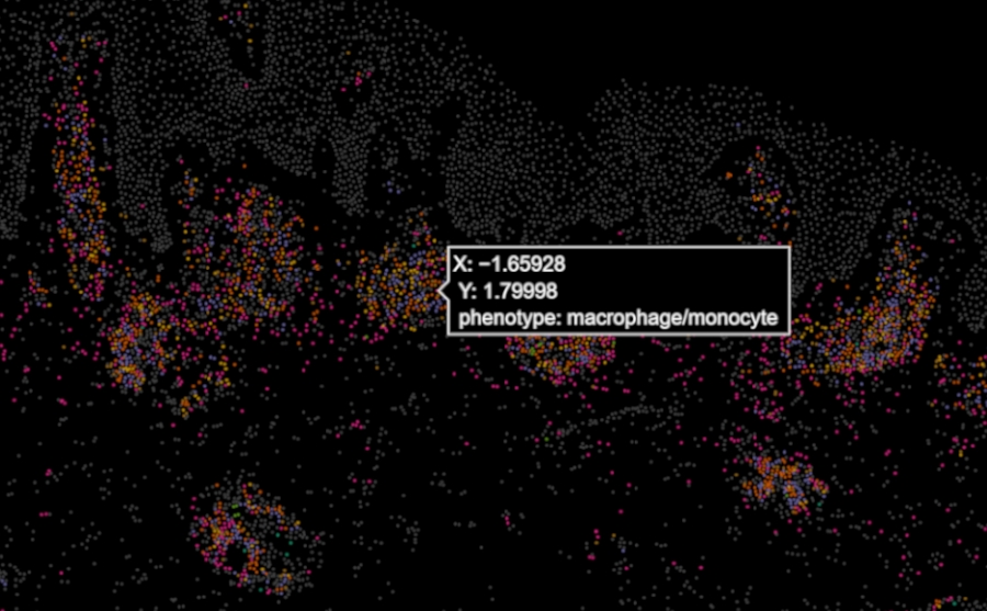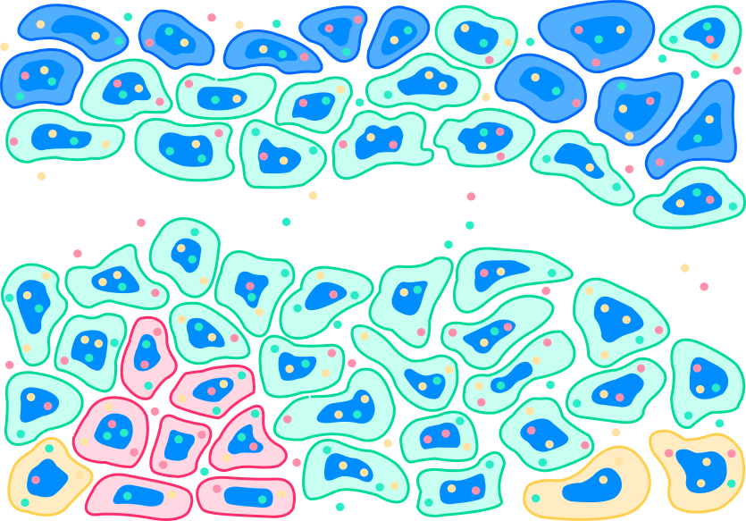
High-plex in situ multiomics at subcellular resolution provides scalable cell atlas and spatial insights with a comprehensive selection of targeted, customisable panels.
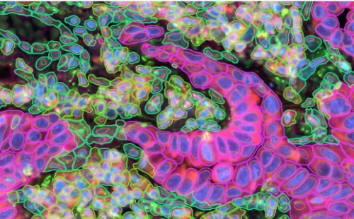
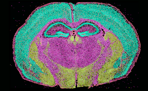
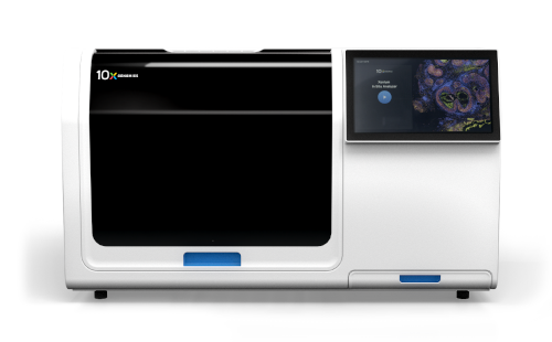








Dual-recognition and ligation-gated chemistry provides high specificity, minimising off-target amplification, increasing the signal to noise and enabling detection of SNPs, fusion transcripts and splice variants.
Short recognition sites provide robust performance in low quality RNA in FFPE embedded samples. Panels of probes can be designed for tissue, pathway and species specific studies with spatial context.




We take care of the complete process from sample preparation to data analysis, with robust QA procedures and complete traceability of your precious samples.
Propath offers pre-developed panels for a range of tissues and can support the design of panel spike in or full panel customisation to ensure sufficient coverage of the target biological systems and intended bioinformatic analyses.
Focus panels can accommodate up to 480 RNA targets and can include a panel of up to 27 protein targets.
In situ imaging is the direct detection of reporter probes at sub-cellular resolution on tissue section. Probes can be hybridised to RNA or protein molecules and identified
through serial cycles of imaging to build in situ barcodes at the site of probe hybridisation. This allows molecules to be identified in space, assigned to cells and localised
to cellular compartments, all while preserving tissue context.
In situ imaging refers to direct, on-slide imaging of reporters, in contrast to workflows with sequencing based readouts that required mapping data back to the slide via spatial reporters such as with Visium or GeoMx
Unlike sequencing‑based platforms, Xenium uses imaging‑based detection of RNA transcripts directly within the tissue, avoiding dissociation or RNA capture onto
arrays. This enables true single‑cell spatial resolution and accurate mapping of gene expression to individual cells, rather than aggregated signals from multi‑cell regions.
Both paraffin embedded, formalin fixed (FFPE) and fresh-frozen (FF) samples are supported. Propath recommended FFPE where possible.
The imaging area is 10.45mm x 22.45mm
Yes, Propath can support the design of panel spike ins and fully custom panels. up to 100 custom RNA targets can be added to any panel, and de novo panels can be
designed with up to 480 targets.
Factors including sample type, sample number, scientific question and study phase
Propath offers a complete range of bioinformatics packages from pre-developed to fully custom solutions, including multi-omic integration with multiplex fluorescence, scRNAseq and more
Our experience across a range of platforms for spatial biology allows us to recommend the most appropriate platform for each research project.
We understand the strengths and limitations of each platform – and how to get the best out of them.