As a spatial biology specialist, Propath understands the challenges of producing reliable and reproducible data from complex images.
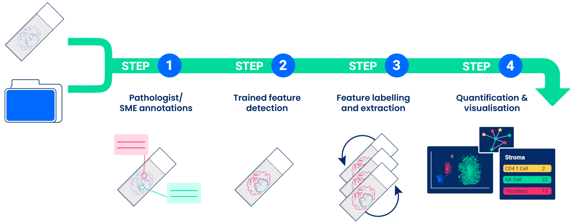
Propath's service for image analysis integrates image management, quantitative quality assessment, spatial phenotyping, and biologically grounded reporting within a validated, reproducible workflow.
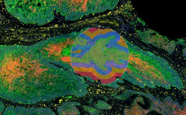
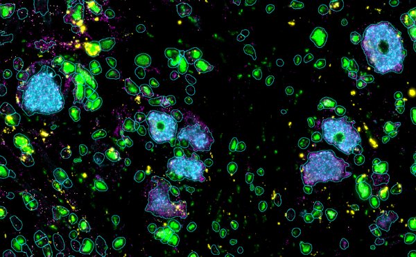
Combining advanced analytics with domain expertise, Propath enables sponsors to extract clear, reproducible meaning from complex tissue data, translating images to impact.
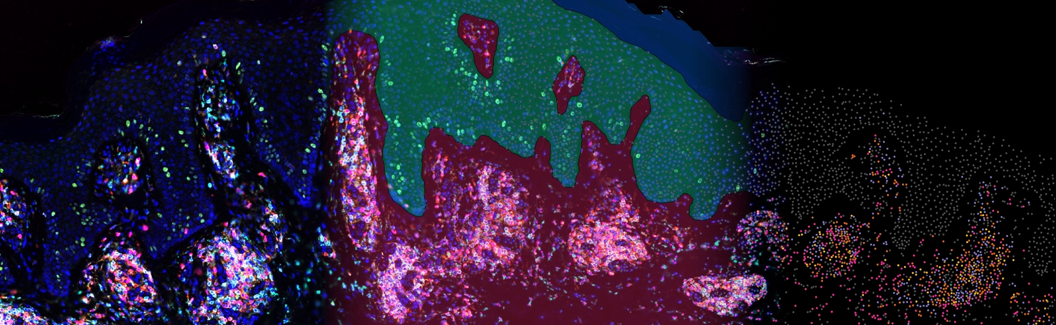
Propath's image management service provides data storage and transfer, image viewing and collaboration tools with no downloads required. Supporting a wide range of image formats and stacking of multiple imaging modalities, analysis overlays and tools for annotation, ensure data is securely accessible on demand.

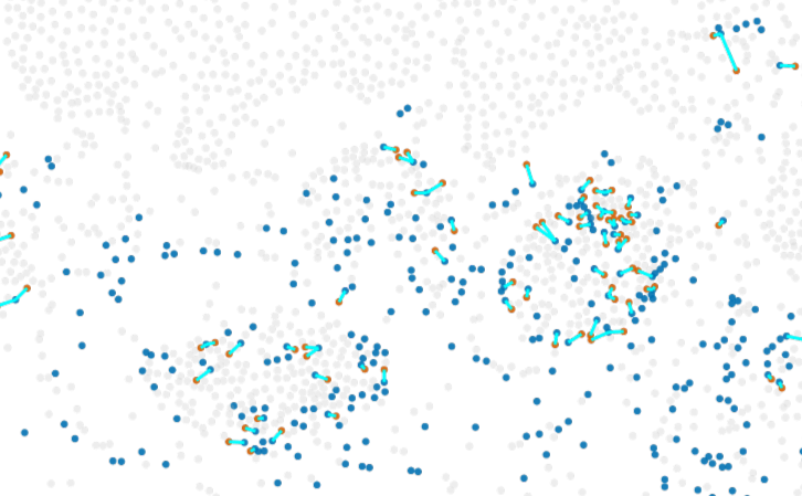
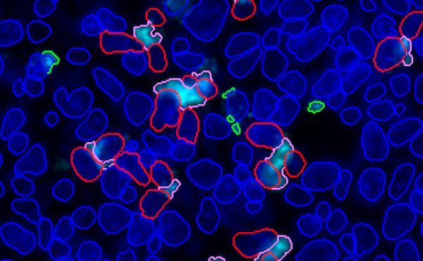
T-cell to macrophage nearest neighbour analysis
Propath's platform for quantitative image analysis and bioinformatics has been designed and validated to be fit-for-purpose for use on clinical trial data.
Our controlled environment ensures analyses are conducted according to the principles of informed consent, data integrity, confidentiality and scientific rigour to maintain GCP compliance from sample receipt to data delivery.
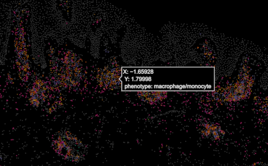
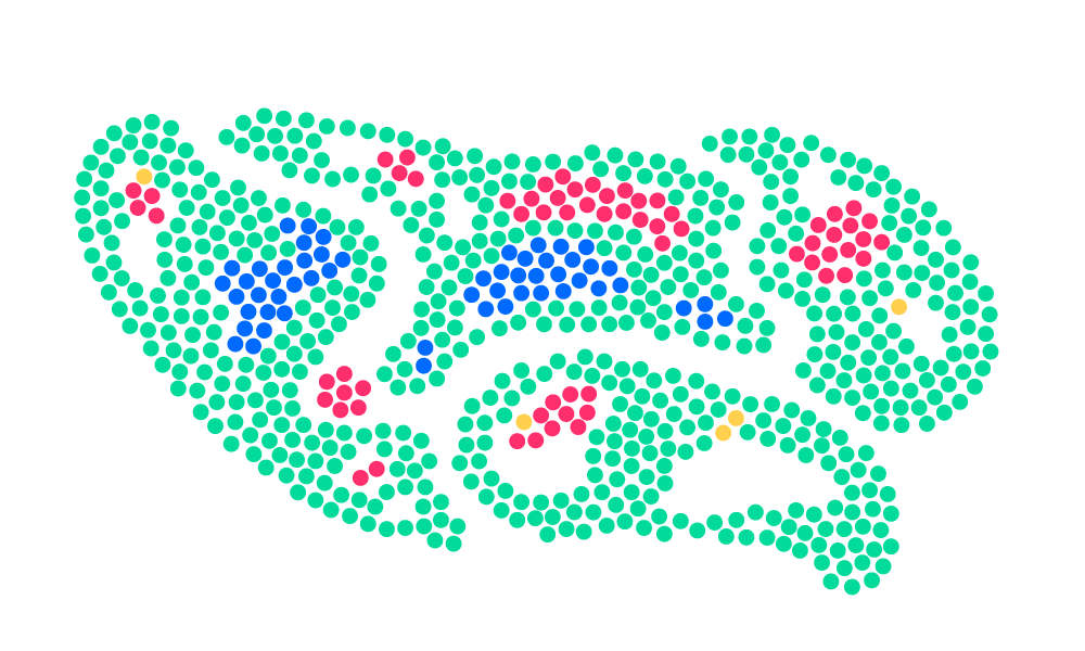
Whether sponsors enter our pathway with their own images or Propath generated images, Propath seamlessly translates qualitative images into quantitative data for statistical analysis and visualisation. In collaboration with pathologists or subject matter experts, algorithms are trained to extract and quantify features, providing robust and reproducible results.
Our experience across a range of platforms for spatial biology allows us to recommend the most appropriate platform for each research project.
We understand the strengths and limitations of each platform – and how to get the best out of them.